本文是一篇医学论文,医学可分为现代医学(即通常说的西医学)和传统医学(包括中(汉)医、藏医、蒙医、维医、朝医、彝医、壮医、苗医、傣医等)多种医学体系。不同地区和民族都有相应的一些医学体系,宗旨和目的不相同。印度传统医学系统也被认为很发达。(以上内容来自百度百科)今天为大家推荐一篇医学论文,供大家参考。
1 Background
Diabetes mellitus is a chronic metabolic disorder that has symptoms that arecharacterized by hyperglycemia, which leads over time to serious damage to the heart,blood vessels, eyes, kidneys, and nerves. Most frequently occur complications ofdiabetes include heart attack, stroke, kidney failure, vision loss, nerve damage andsevere foot ulcers which lead to leg amputation (because of infected, non-healing footulcers)[1]. Diabetic vascular complications are the major contributors to diabetes-associated morbidity and mortality[2]. Previous studies showed that as a result ofdiabetes complication, a lower limb is lost every 30 seconds[3, 4].Peripheral artery disease is a narrowing or occlusion of the peripheral arteries,which results in a gradual reduction of blood supply to the stomach, arms, head, andlower limbs, which is the most common site of peripheral artery disease. In patientswith peripheral arterial disease, compared to non-diabetic patients, diabetic patientshave worse progress and poorer prognosis, leading to a high rate of amputation anddeath[5]. The most advanced, severe symptoms of peripheral artery disease is criticallimb ischemia (CLI), a condition in which limb perfusion is strictly restricted, andcharacterized by pain during rest and skin ulcerations or gangrene of the extremities[6].CLI is also one of the most severe complication of diabetes mellitus. The risk ofdeveloping CLI is two to four-times higher in patients who have diabetes mellitus thanin patients without diabetes mellitus. CLI also progresses more rapidly in patients withdiabetes mellitus[2]. The 6-month major amputation rate and 1-year mortality rate indiabetic CLI patients is 10% to 30% and 25%, respectively; while the 5-year mortalityrate is as high as 30%[7, 8].
1.1 Pathophysiology of Critical Limb Ischemia
The pathophysiology of CLI is a chronic and complex process that affects themacrovascular and microvascular systems. Increased severity of ischemia is associatedwith worse outcomes, such as amputation and death. Severe critical limb ischemia isoften the result of the blockage of the arteries that supply blood to lower limbs[9].The first response of our body against ischemia is to induce angiogenesis, orcapillary sprouting, as well as arteriogenesis, thereby promoting the enlargement of pre-existing collateral blood vessels to increase blood supply to the critically ischemic limb.These responses fail to supply the necessary amount of blood flow and oxygen to theischemic limb, causing CLI patients has vasodilated and insensitive arterioles to pro-vasodilatory stimuli. Furthermore, blood vessels of CLI patients have decreased wallthickness, decreased cross-sectional area, and decreased wall-to-lumen ratio comparedwith normal person[9]. In diabetes, there are also some disturbance in the vessel wallthrough promotion of vascular inflammation, endothelial cell dysfunction, andabnormalities in smooth muscle cells and macrophage. Such vascular abnormalities thatcause CLI in diabetes patients severity increases with worsening blood glucose andduration of diabetes[10].
............
1.2 Roles of Platelet-Derived Growth Factor-BB (PDGF-BB)and Vascular Endothelial Growth Factor (VEGF) in angiogenesis
Therapeutic angiogenesis involves the use of angiogenic growth factors to growblood vessels, improve blood flow, and increase tissue perfusion in the ischemicextremities. The neovascularization process in ischemic tissue is complex and involvesa local increase in growth factors to promote the new blood vessel growth andimmunomodulation of tissue repair and regeneration cell activity. Firstly, newcapillaries are sprouting from post capillary venules, it is stimulated mainly by tissuehypoxia via the accumulation of hypoxia-inducible factor (HIF)-1 protein. HIF-1 -1, ANG2, etc. And then the process ofmaturation seems to involve the release of FGF, PDGF and VEGF[11].In vitro, various growth factors may promote wound healing. Of the numerouscytoactive factors investigated, platelet-derived growth factor-BB (PDGF-BB) is theonly currently approved growth factor therapy for enhancing diabetic wound healing bystimulate proangiogenic factors, stimulate endothelial cell migration, proliferation andincrease wound vascularity[19].Various studies identified that expression of growth factors such as VEGF andPDGF are critically important in the formation of collateral vessels in response toischemia. But in hyperglycemia, decreased VEGF and PDGF expression in theperipheral limbs of diabetic animals has been reported. Although the underlyingmechanism of reduction of VEGF and PDGF expression in diabetes is not clear,however poor collateral vessel formation during diabetes-induced ischemia might beassociated to the lack of production and action of growth factors such as VEGF andPDGF[20]. Previous studies also have shown that PDGF-BB co-delivery significantlyincreased VEGF-induced vascular density and prevented the appearance of aberrantstructures, yielding a network of homogenous, mature capillaries. Thus, PDGF-BBmediated vascular maturation represents a complementary target to both normalize andpotentiate VEGF-induced angiogenesis, with promising implications to significantlyimprove the efficacy of therapeutic angiogenesis strategies[21].
........
2 Effect of salidroside on promoting angiogenic factors inskeletal muscle cells in high glucose condition
Previous studies have found that skeletal muscle cells played important roles inangiogenesis because they could expressed and secreted angiogenic factors viaautocrine or paracrine mechanisms[16, 39]. To reveal the effect of hyperglycemia on theparacrine functions of the skeletal muscle cells, we first cultured C2C12 myoblast cellsin low glucose and high glucose conditions, and compared angiogenic factorsexpression levels.On the other hand, previous studies had also shown that PHD family membersplayed important roles in angiogenesis as they regulated the expression of a number ofangiogenic factors[40]. Thus, we also examined the effect of hyperglycemia on theexpression levels of PHD3 in skeletal muscle cells, and its relation with the expressionlevels of angiogenic factors.Salidroside was used by many people to treat high-altitude sickness due to itspharmacological effect to resist hypoxia[26]. Targeting angiogenic factors to grow bloodvessels, improve blood flow, and increase tissue perfusion in the ischemic extremitiesrepresents a promising therapeutic strategy for CLI in diabetes. Thus, we nextinvestigated the efficacy of salidroside on promoting angiogenic factors in high glucosecondition by evaluating its mRNA and protein expression levels.
2.1 Materials
① Phosphate buffer saline (PBS), pH 7.2 - 7.4One packet of solid PBS was added to 1 L ddH2O. The PBS was then sterilizedin an autoclave for 30 minutes at 120°C. The solution was placed in the aseptic cellculture room and exposed to ultraviolet (UV) overnight.② Complete culture medium (10%)DMEM was supplemented with 10% Fetal Bovine Serum and 1% ofpenicillin/streptomycin.③ Starvation culture medium (no FBS)100 U/ml penicillin/streptomycin was added to DMEM at a 1:100 ratio.④ Glucose 25 mmol/L (high glucose)D-Glucose molecular weight = 180.2 g. PBS was used to dilute solid D-glucose.To make high glucose condition, 9.01 g solid D-Glucose was added to 10 ml PBSSolution. After glucose was dissolved, then it was filtered and placed in the aseptic cellculture room.
............
2.2 Method
1) Before cells were removed from the storage, the complete culture medium waspre-warmed in 37°C water bath.2) The aseptic laminar air flow was prepared by lighting the burner.3) 75% alcohol was used to clean both our hands and aseptic laminar air flow, andto kill off germs and bacteria, and to reduce pollution. New flask or dish plate that weused for cell culture were sterilized first before placing them into the aseptic laminar airflow.4) Sterile pipettes were burned and allowed to cool before starting the experiments.5) Cryopreserved C2C12 cells were taken out from liquid nitrogen canister, andthey were thawed in 37°C water within 1 – 2 minutes.6) The cells that have been thawed was gently added into 15 ml centrifuge tube,then 5 ml complete culture medium was added, mixed, and centrifuged at 1000 rpm in 5minutes. The supernatant was discarded.
..........
3 EFFECT OF SALIDROSIDE ON SKELETAL MUSCLE CELLMOBILITY AND PROLIFERATIONN..........43
3.1 MATERIALS....... 43
3.2 METHOD............ 46
3.3 RESULTS ............ 49
3.3.1 muscle cell migration under high glucose condition.........49
3.3.2 cell proliferation under high glucose condition.......50
3.4 DISCUSSION AND CONCLUSION........ 51
4 OVEREXPRESSION PHD3 INHIBITED THE EFFECT OFSALIDROSIDE ON PROMOTING ANGIOGENESIS..............53
4.1 MATERIALS....... 53
4.2 METHOD............ 58
4.3 RESULTS ............ 70
4.4 DISCUSSION AND CONCLUSION ....... 72
5 EFFECT OF CONDITIONED MEDIUM OF SKELETALMUSCLE CELL SECRETOME.............. 73
5.1 MATERIAL......... 73
5.2 METHOD............ 76
5.3 RESULTS............ 81#p#分页标题#e#
5.4 DISCUSSION AND CONCLUSION ....... 86
7 Effect of salidroside on critical limb ischemia ofstreptozotocin-induced diabetic mice model
When cells were exposed to ischemic stress, then to meet the shortage of oxygenand nutrition supply, the formation of new blood vessels will be occurred. The fact thatin our last experiment that salidroside could enhancing angiogenic factors in vitro inhigh glucose condition, we would like to investigate whether salidroside could promoteneovascularization in ischemic tissue of diabetic mice model. Animal models played animportant role in helping as both guide practice and trials of cell-therapy for CLI. Weexamined that the therapeutic administration of salidroside would promote peripheralangiogenesis in a mouse model of CLI in diabetic so as to reestablish blood flow to theaffected limb. The C57BL/6 mouse strain was used as a model for studies of diet-induced obesity and diabetes[54]. There are several different murine hind limb ischemiamodels to test angiogenesis via cell based therapies. In this study, we testedangiogenesis via unilateral femoral artery ligation.
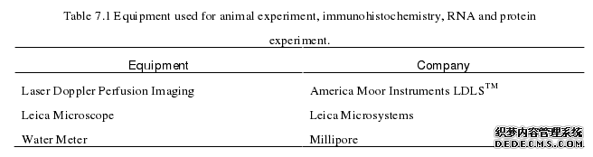
7.1 Materials
Male C57BL/J mice model, inbred strain, 7 weeks old were purchased fromThird Military Medical University, Daping Animal Research Center. Animal studieswere carried out in the Third Military Medical University, and approved by theLaboratory Animal Welfare and Ethics Committee of the Third Military MedicalUniversity. All surgeries were performed under ketamine/xylazine anesthesia(intraperitoneal administration, 80 mg/kg body weight and 50 mg/kg body weight,respectively), and all efforts were made to minimize suffering.
..........
Conclusion
Diabetic patients are at high risk for critical limb ischemia. The incidence andseverity of critical limb ischemia in diabetes is increased approximately twofold tofourfold[2]. In recent years, many research conducted preclinical and clinical trials forthe treatment in critical limb ischemia in diabetes. However, none of them haveobtained effective therapeutic treatment. Previous research reported that therapeuticangiogenesis had been considered as current gold standard for treating critical limbischemia in diabetes, therefore, many scientists are now searching for new targets andnew treatments for therapeutic angiogenesis, such as gene therapy or cell therapy[7].Salidroside is an active ingredient isolated from Rhodiola, a well-known herb usedin Eastern Europe and Asia to relieve high altitude sickness for hundreds of years. It isbelieved to possess various pharmacological activities, such as resisting anoxia, anti-oxidant, anti-inflammatory, anti-apoptotic, anti-cancer, anti-depressive, anti-aging,neuroprotective, hepatoprotective and cardioprotective effects[26-30]. In addition, studyhas reported that salidroside could promote the stability of HIF-endothelial cells, while promoting the expression of angiogenic factors anderythropoietin[58]. Another research also reported that salidroside promotes themigration and proliferation of bone marrow progenitor cells, promotes thedifferentiation of endothelial precursor cells, and promotes the phosphorylation of Akt,thereby facilitating the involvement of bone marrow progenitor cells in the formation ofblood vessel[59]. Our previous study has also reported inhibition of PHD3 by salidrosidepromotes functional angiogenesis through paracrine signaling of skeletal muscle cell[17].The neovascularization process in ischemic tissue is very complex, it requires alocal increase of angiogenic factors, tissue protection factors and regeneration cellularactivity. The new sprouting angiogenesis occurred in several stages. Firstly, in ischemictissue, induced by hypoxia, the activation of HIF-1 expression leads to angiogenicfactors, such as VEGF, ANG2, neuropilin-1, etc., activate receptors on endothelial cellspresent in pre-existing blood vessels.
..........
References (abbreviated)

